Massage

Definition
Massage is the scientific manipulation of soft tissues of the
body with the palmar aspect of hands(s) and or fingers.

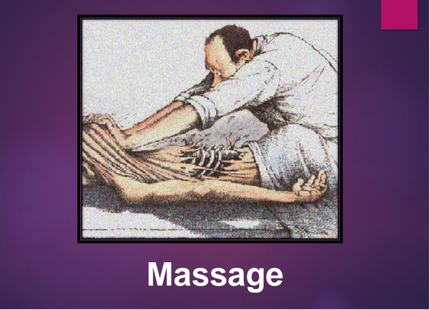
 Massage is a tern applied to certain manipulation of the soft
tissues.

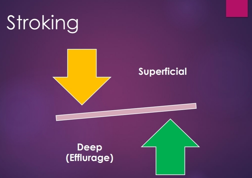
Superficial stroking
 the rhythmic movement of hand or parts of hand, over
the skin the lightest amount of pressure in order to
obtain sensory stimulation.
 Strokes can be applied from proximal to distal part or
vice versa. (Centrifugal of Centripetal) .
 Every massage should begin and end with strokes.
 It can also be used between every other technique.
 It is best performed with palm of hand or pulp of fingers.

 This “just ‘’ touch no pressure contact should be
maintained throughout the stroke.
 The hands should work in alternate motions, so that there
is no loss of contact with patients Skin.
 At a time only one hand should slide over the pt’s skin.
 The stroke should end with a smooth lift of hand.
 Slower strokes (12-15/min) are more sedative.
 Faster strokes (30-40/min.) are stimulating in nature. PHYSIOLOGIC EFFECT
 Soothing & sedative
 Stimulates cutaneous touch receptors.
 Indirectly improves circulation by activating the axon
reflex.
 Facilitates contraction of muscles. effects. THERAPEUTIC EFFECT.
 Reduces anxiety, tension & psychological stress.
 Decrees hyper sensitivity
 Fast stroking elicits contraction in hypotonic
muscles. *Deep stroking / Effleurage
 It is the movement of the palmar aspect of hand over the external
surface of the body With constant moderate pressure, in the
direction of venous and lymphatic drainage.
 Each stroke begins from the distal end of the segment and is
completed at the proximal end usually at the site of a group of
lymph nodes.
 Direction of movement of hand is always from distal to proximal.
 Contact and Continuity must be even throughout the stroke.
 Depending on area, one or both hands may be used.  Pressure is applied by transfer of body weight to the
subject skin through the upper extremity of therapist.
 Return stoke is usually superficial.
 Rate of stroking is usually slow (10-12/min) to allow
refilling of venous & lymphatic Channels.
 In babies with “C” grasp effleurage is done from
proximal to distal. Therapeutic uses
 Removal of edema.
 Removal of metabolites & inflammatory
products.

Pressure Manipulation
Kneading
• Palmar, Digital, Ironing.
Petrissage
•Picking-Up, Wringing, and Skin rolling.
Friction
Circular, Transverse

Kneading
 It is a pressure manipulation
 It is a technique in which tissues is pressed down on to the
underlying structures.
 Pressure is applied in a circular way along the long axis of
underlying bone.
 The Pressure is Increase and Decrease in gradual manners.
 Hands are placed over the skin and tissues are
compressed against the bone and hands are moved in a
circular direction.

 Several small concentric circles are performed to the body surface
and each circle overlaps the previous one.
 ½ the circle Pr. is Increase and ½ it is Decrease.
 Thus each circle has 2 phases.
 (i) Phase of compression
 (ii) Phase of relaxation
 Increase Pr. should always be proximal to distal.
 Kneading can be applied with part of fingers, thumb or palm of
one or both hands.

Palmar Kneading
 Performed with whole of palmar or with heel of hand.
 Usually performed over larger areas such as thigh, calf, arm etc.
 Both the palms are placed on opposite aspects of limb segment (medial and
lateral)
 Fingers & thumb not in a contact with the skin.
 Both hands should perform co-ordinate circular movement, in opposite
directions.
 ½ Cycle pr Increase and ½ cycle it decreases.
 Slow rates of kneading allow deeper penetration.

Digital Kneading (Finger / Thumb)
Finger kneading
 palmar aspect of whole finger or part of it may be used.
 Whole finger kneading, finger pad kneading & fingertip kneading
can be used.
 One finger or 2-3 fingers may be used. To increase contact area.
 Thumb & little finger should have no contact the skin.

Thumb Kneading
 The tip of one or both the thumbs are used depending on the
side of area .
 One thumb may be placed over the other to reinforce the
movement.
 Thumb pad and thumb tip kneading techniques are used.
 Thumb pad – smaller muscular areas such as thenar eminence.
 Thumb tip- Narrow area such as interosseous spaces.

Reinforced kneading / Ironing
 Utilizes both the hands for kneading
 Used when greater depth is required.,
 Most commonly used over back.
 Hands placed over each other on the skin.
 Elbow of therapist in extension.
 Intermittent circular Pr. is transmitted to the body.

Petrissage or working
 Derived form French word “Petrir’’ meaning to “knead’’.
 The direction of application of Pr. differentiates petrissage from
Kneading
 Kneading exerts vertical compression of soft tissues over underlying
bone and petrissage involves picking up movement of soft tissue with a
lateral compression.
 Intermittent Pr. is applied at right angles to the long axis of bone.
 Picking up, skin rolling & wringing are the petrissage techniques.

Picking Up
 Involves lifting of the tissue up at right angles to the
underlying bone, squeezing and Releasing it.
 The web space between thumb and index finger lies across
the central line of the muscle bulk and skin to be lifted.
 The thumb and thenar eminence are placed on one side and
index, middle finger placed over the other side of the central
line.

 Transfer of body weight to skin through upper extremity to apply compressions
initiates the technique.
 Same time grasp of hand is tightened along with little extension of wrist.

Skin Rolling
 Involves lifting and stretching of the skin between the thumb and the fingers as
well as moving the skin over the subcutaneous tissue.
 Therapist lifts up and moves the skin and superficial fascia with both the hands
keeping a roll of the skin raised continuously ahead of the moving thumb.
 Thumb is abducted in such a way that the tip of thumb and index fingers of both
hands touch each other maintaining a full palmar contact with the skin.
 Pull the finger backwards with sufficient pressure so that skin is pulled up.
 At the same time thumb is adducted and opposed with downward pressure over
the underlying skin.

 The coordinated motion of thumb and fingers lifts off a roll of skin from
the underlying structures.
 Palm is gradually lifted off the skin so that lifted roll of skin remains
between tip of fingers and thumb.
 Move the thumb forward to roll the lifted skin against the fingers.
 Main effect of this technique is to stretch the cutaneous and subcutaneous
tissue and induce relaxation.
 Usually performed over the back.
Skin Rolling.

Muscle Rolling
 Applied over arm, calf thigh muscles.
 Whole muscle is lifted between the tip of thumb and fingers,
using similar grasp
 Alternately apply and release pressures with thumb and fingers
which rolls muscle fibers from side to side.

Wringing
 Grasp and placement of hand similar to picking up.
 Both hands are used in opposite aspects of the limb.
 Lifting up the skin is done and both hands move in opposite
directions.(Forward & backward)
 Useful for mobilization of adherent skin.

Physiologic & Therapeutic uses
(Kneading & petrissage)
 Mainly break down of adhesions.
 Reduce edema.
 Increase mobility of adherent structures.

Friction or Rubbing
 It consists of small range oscillatory movement which is applied
to the deeper structures Pressure by thumb or fingers.
 According to direction of movement it grouped as.
 (i) Circular Friction
 (ii)Transverse friction.

Circular friction
 Advocated by wood in1974. Resembles digital kneading.
 Only difference is continuous Pressure is applied during whole
procedure no phase of Relaxation.
 Movement in circular direction. Increase Pressure when superficial
Structures become relaxed.
 Applied around localized area (joints, muscle attachments fibrocytic
nodules.)
 Used in case when nerve trunk is imbedded in consolidated edema
fluid. Localized effects on muscles are in prolonged state of tension.

Transverse friction
 Movement is transverse (i.e.) across the long axis of the structure to be
treated.
 Performed with tip of the thumb, index or middle finger, which can be
reinforced, 2 or 3 fingers.
 Ligaments to be treated should be in taut position.
 Muscles to be treated should be in relaxed position.
 It is a painful procedure and patient is to be informed about it priorly.

Physiologic and therapeutic effects
❖ Breaks down intra-fibrillary adhesions.
❖ Smoothens the rough gliding surfaces.
❖ Ensures pain free mobility.
❖ Useful in traumatic muscular lesions, tendonitis, tenosynovitis and
ligament sprain.
❖ Much useful in localized pain (trigger points).

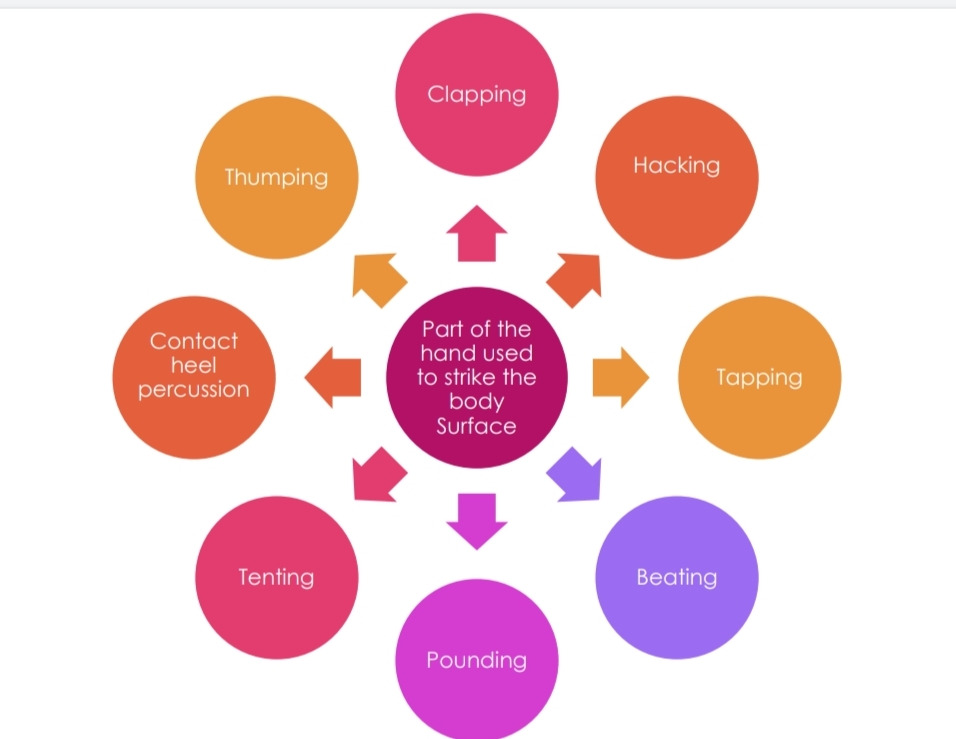
Percussion or Tapotement
 French word which means striking of two objects against each
other.
 Utilizes controlled movement of wrist and forearm to strike the
patients body surface Rhythmically.
 Mild blows are applied with various pressure and in different
manners.

Clapping or Slapping
 Used in management of chronic respiratory disorders which
leads to sputum retention
 Slightly cupped hands strike the chest wall alternately in
predetermined rate.
 Fingers and thumb are adducted and M.P. joints of fingers are
slightly flexed in position.
 Palm should not come in contact with patient’s body during
striking.

 No movement at elbow, only wrist flexion and extension should be
done rapidly.
 Hand should create air cushion between the hand and the chest
wall on impact.
 Performed during both inspiration and expiration.
 Manual percussion normally 100 – 480 times / Min.
 Clapping is performed over chest wall, with a blanket or towel
covering it.
Clapping or Slapping.

Hacking
 The ulnar border of medial 3 fingers is used to strike the skin.
 This produces a peculiar sound
 Alternate supination and pronation of forearm combined with
radial deviation of the wrist respectively produce hacking.
 Applied over larger areas such as back and thigh etc.

Tapping
 Useful when intermittent touch and pressures are to be applied over a
small area.
 Only the pulp of fingers strikes the body part.
 One of both hands may be used.
 Alternate flexion and extension of the MCP joints produce the tapping.
 No movement should be at wrist and elbow.
 Commonly used over face, neck and other smaller areas.
 Conveniently used on children.

Beating
 Anterior aspect of the clenched fist strikes the body part.
 In making fist, fingers are flexed at MCP and PIP joints but DIP is
kept extended to produce a flat surface, which is used for beating
 Beating is produced by alternate flexion and extension of wrist.
 No movement at elbow.
 Used over back, thigh and other fleshy and broad area of body.

Pounding
 Fingers are flexed at all the joints to make a fist.
 Thumb rests over the index finger.
 Supination and pronation of forearm combined.
 Ulnar and radial deviation of wrist respectively produces the pounding
 Used over back, thigh and other fleshy and broad areas of the body.
 Ulnar border of clenched fist strikes the body.

Tenting
 It is a modification of clapping.
 Here concavity is produced between the index and the ring finger with the
middle finger slightly elevated and placed over them.
 For loosening secretions in the smaller chest of newborn or a premature infant.

Contact Heel percussion
 It is a modification of clapping.
 Concavity is produced between thenar and hypothenar
eminences.

Thumping
 Apply pressure over the body surface with dorsal aspect of clenched fist.
 Usually applied over back and chest.
 Helps in increase exhalation.
 Helps in coughing up material from bronchial emphysema.

Vibration
 Involves constant touch of therapist hand and application of rapid intermittent
pressure with out changing the position of hand.
 Usually used over chest wall.
 Can be produced by one fingertip or palm.
 The therapist should produce isometric contraction of all the muscles of upper
extremity which are transmitted to the patient’s body surface through his hands.
 This produces oscillatory movements of his hand in upward and downward
direction and
 Transmits the mechanical energy to the patient’s chest.

Shaking
 Oscillation is coarse compared to vibration.
 Fore arm in midprone position.
 Oscillation occurs in side ways movement
 Patients in supine lying, Place both hands on each side of anterior Chest
wall or antero posteriorly on same side.
 Patient’s in side lying , place both hands on the upper lateral chest wall
or antero posteriorly on the upper side chest wall .

 Shaking is done during expiratory phase.
 Therapist tends to produce upward and down ward movement of upper extremity.
 This shakes chest wall vigorously .
 While shaking is done for extremities , they should be raised to encourage venous
and lymphatic return .

MMT=manual muscles testing

1) INTRODUCTION:
 Manual muscle testing is used to determine the extent and degree of muscular weakness resulting from disease, injury or disuse. The records obtained from these tests provide a base for planning therapeutic procedures and periodic re-testing. Muscle testing is an important tool for all members of health team dealing with physical residuals of disability.

 Muscularstrength:

The maximal amount of tension or force that a muscle or muscle group can voluntarily exert in a maximal effort; when type of muscle contraction, limb velocity and joint angle are specified.


 Muscularendurance:
The ability of a muscle or a muscle group to perform repeated contractions
against resistance or maintain an isometric contraction for a period of time.


 Musclepower:
 Power is defined as the generate as much force as fast as possible.  Power does require strength and speed to develop force quickly.
 POWER = strength speed.

 TYPES OF MUSCLE WORK:

  1. Isometric contraction: Tension is developed in the muscle but no movement occurs; the origin and insertion of the muscle do not change their positions and hence, the muscle length does not change.
  2. Isotonic contraction: The muscle develops constant tension against a load or resistance. There are two types:
    a) Concentric contraction: Tension is developed in the muscle and the origin and insertion of the muscle move closer together; so the muscle shortens.
    b) Eccentric contraction: Tension is developed in the muscle and the origin and insertion of the muscle move further a part; so the muscle lengthen.
  3.  RANGE OF MUSCLE WORK:
    The full range in which a muscle work refers to the muscle, changing from a position of full stretch and contracting to a position of maximal shortening. The full range is divided into three parts:
  4. Outer range: From a position where the muscle is fully stretched to a position halfway through the full range of motion.
  5. Inner range: From a position halfway through the full range of motion to a position where the muscle is fully shortened.
  6. Middle range: The portion of the full range between the mid-point of the outer range and the midpoint of the inner range.
  7. GROUP OF MUSCLE ACTION:
  8. Prime mover or agonist:
    A muscle or muscle group that makes the major contribution to movement at the joint.
  9. Antagonist:
    A muscle or a muscle group that has an opposite action to the prime movers. The antagonist relaxes as the agonist moves the part through a range of motion.
  10. Synergist:
    A muscle that contracts and works along with the agonist to produce the desired movement. There are three types of synergists:
    a) Neutralizing or counter-acting synergist b) Conjoint synergist
    c) Stabilizing or fixating synergist
    a) Neutralizing or counter-acting synergists:
    Muscles contract to prevent any unwanted movement produced by the prime mover. For example, when the long finger flexors contract to produce finger flexion, the wrist extensors contract to prevent wrist flexion from occurring. b) Conjoint synergists:
    Two or more muscles work together to produce the desired movement. For example, wrist extension is produced by contraction of extensor carpi radialis longus, carpi radialis brevis and extensor carpi ulnaris muscles. If the extensor carpi radialis longus or brevis contracts alone, the wrist extends and radially deviates, while if the extensor carpi ulnaris contracts alone, the wrist extends and ulnarly deviates. When the muscles contract as a group, the deviation action is cancelled, and the common action occurs. c) Stabilizing or fixating synergists:
    These muscles prevent or control the movement at joints proximal to the moving joint to provide a fixed or stable base, from which the distal moving segment can effectively work. For example, if the elbow flexors contract to lift an object off a table anterior to the body, the muscles of the scapula and gleno-humeral (shoulder) joint must contract to either allow slow controlled movement or no movement to occur at the scapula and gleno-humeral joint to provide the elbow flexors with a fixed origin from which to pull. If the scapular muscles do not contract, the object cannot be lifted as the elbow flexors will act to pull the shoulder girdle downward. 2) DEFINITION OF MMT:
  11.  Manual muscle test (MMT) is a procedure for the evaluation of strength of individual muscle or muscles group, based upon the effective performance of a movement in relation to the forces of gravity or Manual Resistance through the available Range of motion (ROM).
     BASIC COMPONENTS OF MOTOR EXAMINATION:
    i. Nutrition or Bulk of muscle
    ii. Tone
    iii. Reflexes
    iv. Range of motion (ROM)
    v. Manual muscle test (MMT)
    vi. Functional Assessment
    *Important of the Sequence 
    Individual versus group muscle test:
    Muscles with a common action or actions may be tested as a group or each muscle may be tested individually. For example, flexor carpi ulnaris and flexor carpi radialis muscles may be tested together as a group in wrist flexion. Flexor carpi ulnaris may be tested more specifically in the action of wrist flexion with ulnar deviation. On the other hand, Flexor carpi radialis longus and brevis may be tested more specifically in the action of wrist flexion with radial deviation. 3) Purposes and uses of MMT:
     CLINICAL USES :
    i. The severity of problem can be understand. (It is diagnostic Tool)
    ii. We can planning our treatment goals.
    iii. Determine the extend & degree of muscular weakness resulting from disease, injury.
    iv. Correlating muscle picture with in level innervations (myotoms) .
    v. MMT is an Important tool for all the members of the Rehabilitation team.
    vi. Prevents deformities by locating problem areas.
    vii. Help and Evaluate effectiveness of treatment to the therapist.  WHY MMT IS PERFORMED? :
    To get some answers such as:-
    i. Is a particular muscle is normal?
    ii. Is it weak? (How much weak)
    iii. Is it strong enough? (How much strong)
    iv. Is it weak on both the side (bilateral symmetrical)?
    v. Is it weak only on one side (Unilateral)?
    vi. Is proximal muscles are weaker than the proximal one?
    vii. Is there any particular pattern of muscle weakness? 4) GRADES OF MMT:
    i. MRC Scale
    ii. OXFORDScale iii. KENDALL Scale iv. And Others .
  • or – GRADES : 5) PRINCIPLES OF MMT:
    1) Position
    2) Stabilization
    3) Demonstration
    4) Application of Grades
    5) Application of Resistance 6) Checking normal strength 7) Objectivity
    8) Documentation 1) POSITION : PATIENT POSITION:
     Patient is positioned Eliminated or Against gravity. (Patient depend upon testing on muscle or muscles group).
     Do not change patient position repeatedly.
     The patient should be as free as possible from discomfort or pain for the duration of each test. It may be necessary to allow some patients to move or be positioned differently between tests.
     Patient position should be carefully organized so that position changes in a test sequence are minimized. The patient’ s position must permit adequate stabilization of the part or parts being tested by virtue of body weight or with help provided by the examiner. JOINT POSITION:
     The joint position is also changed depend upon their performance.
     Distal part of the joint is moved.
     Place the joint in Antigravity position- Grade 3
     Place the joint in Horizontal position – Grade 4 2) STABILIZATION :
     Patient could stabilizes our self during performed Antigravity position.  The hand placement of the therapist is important.
    HAND PLACEMENT:
    I. PROXIMAL HAND – At Origin of muscle & proximal joint giving stabilization.
    II. DISTAL HAND – Distally offering resistance or Assistance depend upon performance. 3) DEMONSTRATION:
     Demonstrate the desired movement.
     Therapist demonstrate the application of movement or performance to the
    patient.
    4) APPLICATIONS OF GRADES:
     Always start with GRADE 3 (If you start to examine the muscle power, first you should test the grade 3).
     Isolation of muscle could be tested. 5) APPLICATIONS OF RESISTANCE:
     Resistance is applied slowly & gradually.
     Increasing or decreasing manual resistance.
     Increasing length of weight arm.
     Apply presence opposite to the line of pull (Grade 4,5)
     Apply force distally.
     It varies between the persons.
     Use long lever to applied resistance whenever it possible. 6) CHECKING NORMAL STRENGTH:
     Therapist to check the strength of the muscle normal side first. 7) OBJECTIVITY:
     Therapist ability to palpate and observe the tendon or muscle response in very weak muscles. 8) DOCUMENTATION:
     Examiners complete testing documentation or Record first.  This will help for next step of treatment applications.
     And help for checking improvement of treatment. 7) INDICATIONS OF MMT:
    1)Lower Motor Neuron (LMN) Disease.
    2) Some other Neurological (Neuromuscular )disease. Such as,
     Multiple Sclerosis
     Muscular distrophy
     Amyotropic Lateral Sclerosis
     Myasthenia Gravis.
     Guillian – barre syndrome (GBS), etc….
    3) Some Musculoskeletal disorders. 8) CONTRAINDICATIONS OF MMT:
    1) Cerebral Palsy
    2) Cardio vascular disease / Brain injury
    3) Dislocated/ unhealed fracture
    4) Myositis ossifications
    5) Parkinson’s disease
    6) Pain
    7) Inflammation /(inflammatory disease in muscles and or joints)
    8) Severe cardiac & respiratory disease . Cont.
    9) Subluxation joint 10) Hemophelia 11) Osteoporosis 9) PRECAUTION:
    1) Considered contraindications
    2) Do not harm (Be gentle)
    3) Respect pain
    4) Examiner know the available ROM. 5) Follow the principles of procedure 6) Take care of patient comfort
    7) Record accurately.
    8) Extra care taken to giving Resisted Exercise. Cont.
    9) Abdomen surgery or hernia 10) Newly united fracture
    11) Bony ankylosis
    12) Hematoma
    13) If patients take muscle relaxers and or pain medications 14) Prolonged immobilization  ExtracaremustbetakenwhereResistedmovementsmight aggravate the condition:
     Patients with history at risk of having cardiovascular problems.
     Abdominal surgery or herniation of abdominal wall to avoid stress on the
    abdominal wall.
     Fatigue exacerbate the patients condition.
     Patient with extreme debility, for example,
     Malnutrition  Malignancy
    And Severe COPD. 10) LIMITATION OF MMT:
    1) UMN LESIONS :
    Spastic muscle have poor control from higher centers thus its better to go for voluntary control assessment rather than MMT.
    2)RESTICTED ROM DUE TO TCD’S (Transcranial Doppler) :
    TCD’s can give faulty interpretation about MMT, thus in case always mention about the MMT within available range along with Grade.
    3) PRESENCE OF PAIN & SWELLING:
    pain and swelling increases the intra articular tension causing irritation of joint and can affect the MMT result, thus in case always mention about presence of pain along with Grade. 4) TYPES OF CONTRACTION :
    MMT gives idea about Quality of concentric contraction only. (Not Eccentric which is more functional).
    5) UNDERSTANDING OF COMMANDS:
     Paediatric Age group < 5 years  IQ
     Higher functions.
    6) STRENGTH Vs ENDURANCE:
    MMT give knowledge about only the strength and not endurance. 7) Subjectivity (patient) HOOVERS sign
    8)And following methods also Limit the MMT ;
     Showing the Co-ordination
     Showing pictures of gross / patient muscle contraction
     Showing the ability of client to use muscle power
     Showing the how much joint ROM the individual is working through. 11) PROCEDURE:
    1) Explanation & Instruction
    2) Assessment of normal muscle strength
    3) Properly positioned the patient
    4) Stabilization
    5) Substitution movements & Trick movements 6) Conventional methods
    7) Alternating techniques. PREPARATIONS:  The plinth or mat table for testing must be firm to help stabilize the part being tested. The ideal is a hard surface, minimally padded or not padded at all. The hard surface will not allow the trunk or limbs to “sink in. ” Friction of the surface material should be kept to a minimum. When the patient is reasonably mobile a plinth is fine, but its width should not be so narrow that the patient is terrified of falling or sliding off. When the patient is severely paretic, a mat table is the more practical choice. The height of the table should be adjustable to allow the examiner to use proper leverage and body mechanics.  Materials needed include the following:
    • Muscle test documentation forms
    • Pen, pencil, or computer terminal
    • Pillows, towels, pads, and wedges for positioning • Sheets or other draping linen
    • Goniometer
    • Interpreter (if needed)
    • Assistance for turning, moving, or stabilizing the patient • Emergency call system (if no assistant is available)
    • Reference material 1) EXPLANATION & INSTRUCTION:
    The therapist demonstrate and or explains briefly the movement to be performed and or passively moves the patient’s limb through the test movement.
    2) ASSESSMENT OF NORMAL MUSCLE STRENGHT:
    Always assess and record the strength of the unaffected side limb to determined the patient’s normal strength. 3) PROPERLY POSITINED THE PATIENT:
    The patient is positioned to isolate the muscle (or) muscles group to be tested in either gravity eliminated or Against gravity positioned. 3) STABILIZATION:
    I. PROXIMAL HAND – At Origin of muscle & proximal joint giving stabilization.
    II. DISTAL HAND – Distally offering resistance or Assistance depend upon performance.
     The plinth or mat table for testing must be firm to help stabilize the part being tested.  The site of attachment of the muscle origin should be stabilized, so the muscle has a fixed point from which to pull. Substitutions and trick movements are avoided by making use of the following methods:
    a) The patient’s normal muscles: For example, the patient holds the edge of the plinth when hip flexion is tested and uses the scapular muscles when gleno- humeral flexion is performed.
    b) The patient’s body weight: It is used to help fix the proximal parts (shoulder or pelvic girdles) during movement of the distal ones.
    c) The patient’s position: For example, when assessing hip abduction strength in side lying, the patient holds the non-tested limb in hip and knee flexion in order to tilt the pelvis posteriorly and to fix the pelvis and lumbar spine.  d) External forces: They may be applied manually by the therapist or mechanically by devices such as belts and sandbags.
     e) Substitution and trick movements: When muscles are weak or paralyzed, other muscles may take over or gravity may be used to perform movements normally carried out by the weak muscles. 4) CONVENTIONAL METHODS:
     Manual grading of muscle strength is based on three factors:
  • Evidence of contraction: No palpable or observable muscle contraction (grade 0) or a palpable
    or observable muscle contraction with no joint motion (grade 1).
  • Gravity as a resistance: The ability to move the part through the full available range of motion with gravity eliminated (grade 2) or against gravity (grade 3).
  • Amount of manual resistance: The ability to move the part through the full available range of motion against gravity and against moderate manual resistance (grade 4) or maximal manual resistance (grade 5).
  • Adding (+) or (-) to the whole grades: This is needed to denote variation in the range of motion. Movement through less than half of the available range of motion is denoted by a “+” (outer range), while movement through greater than half of the available range of motion is denoted by “-“ (inner range). CONVENTIONAL GRADING: Numerals Letters
    The patient is able to move through:
    Description
    Against gravity tests:
    5
    N (normal)
    The full available ROM against gravity and against maximal manual resistance, with hold at the end of the ROM (for about 3 seconds).
    4
    G (good)
    The full available ROM against gravity and against moderate manual resistance.
    4-
    G – (good -)
    Greater than one half of the available ROM against gravity and against moderate manual resistance.
    3+
    Gravity eliminated tests:
    F + (fair +)
    Less than one half of the available ROM against gravity and against minimal manual resistance.
    3
    F (fair)
    The full available ROM against gravity.
    3-
    F – (fair-)
    Greater than one half of the available ROM against gravity.
    2+
    P + (poor +)
    Less than one half of the available ROM against gravity.
    The patient is able to actively move through:
    2
    P (poor)
    The full available ROM with gravity eliminated.
    2-
    P – (poor -)
    Greater than one half the available ROM with gravity eliminated.
    1+
    T + (trace +)
    Less than one half of the available ROM with gravity eliminated.
    1
    T (trace)
    None of the available ROM with gravity eliminated and there is palpable or observable flicker contraction.
    0
    0 (zero)
    None of the available ROM with gravity eliminated and there is no palpable or observable muscle contraction. SCREENING TEST:
     A screen test is a method used to control muscle strength assessment, avoid unnecessary testing and avoid fatiguing and / or discouraging the patient. The therapist may screen the patient through the information gained from:
  1. The previous assessment of the patient’s active range of motion.
  2. Reading the patient’s chart or previous muscle test result.
  3. Observing the patient while performing functional activities. For example, shaking the patients hand may indicate the strength of grasp (finger flexors).
  4. All muscle testing procedures must begin at a particular grade; this is usually grade “fair”. The patient is instructed to actively move the body part through full range of motion against gravity. Based upon the results of this initial test, the muscle test is either stopped or proceeds.
  5. FACTORS AFFECTING STRENGTH:
    1). Age:
    A decrease in strength occurs with increasing age due to deterioration in muscle mass. Muscle fibers decrease in size and number; there is an increase in connective tissue and fat and the respiratory capacity of the muscle decreases. Strength apparently increases for the first 20 years of life, remains at this level for 5 or 10 years and then gradually decreases throughout the rest of life. The changes in muscular strength by aging are different for different groups of muscles. The progressive decrease in strength is clearer in the forearm flexors and muscles that raise the body (anti-gravity muscles). 2). Sex:
    Males are generally stronger than females. The strength of males increases rapidly from 2 to 19 years of age at a rate similar to weight and more slowly and regularly up to 30 years. After that, it declines at an increased rate to the age of 60 years. The strength of females is found to increase at a more uniform rate from 9 to 19 years and more slowly to 30 years, after which it falls off in a manner similar to males. It has been found that women are more 28 to 30% weaker than men at 40 to 45 years of age. 3). Type of muscle contraction:
    More tension can be developed during an eccentric contraction than during an isometric contraction. The concentric contraction has the smallest tension capability.
    4). Muscle size:
    The larger the cross-sectional area of a muscle, the greater the strength of this muscle will be. When testing a muscle that is small, the therapist would expect less tension to be developed rather than if testing a large, thick muscle. 5). Speed of muscle contraction:
    When a muscle contracts concentrically, the force of contraction decreases as the speed of contraction increases. The patient is instructed to perform each movement during muscle test at a moderate pace.
    6). Previous training effect:
    Strength performance depends up on the ability of the nervous system to activate the muscle mass. Strength may increase as one becomes familiar with the test situation. The therapist must instruct the patient well, giving him an opportunity to move or be passively moved through the test movement at least once before muscle strength is assessed. 7). Joint position:
    It depends on the angle of muscle pull and the length-tension relationship. The tension developed within a muscle depends upon the initial length of the muscle. Regardless of the type of muscle contraction, a muscle contracts with more force when it is stretched than when it is shortened. The greatest amount of tension is developed when the muscle is stretched to the greatest length possible within the body (if the muscle is in full outer range).
    8). Fatigue:
    As the patient fatigues, muscle strength decreases. The therapist determines the strength of muscle using as few repetitions as possible to avoid fatigue.
    The patient’s level of motivation, level of pain, body type, occupation and dominance are other factors that may affect strength.  BREAK TEST:
     Resistance applied at the end of tested range is termed as BREAK TEST.
     For one joint muscle resistance is applied at End of ROM.
     For two joint muscle resistance is applied at Mid Range.
     The isometric hold (break test) shows the muscle to have a higher grade than the make test.
     MAKETEST:
    Resistance is applied throughout the test is called MAKE TEST.  INDICATIONS OF BREAK TEST:
  6. When movement is contraindicated
  7. When there is pain in movement
  8. When we have to assess the quality of strength and not the quantity?. INTRUMENTATION:
     Instrument chosen to assess muscle strength depends on the degree of accuracy required in the measurement. HAND HELD DYNAMOMETER:
    This Device operate on principle of compression. Application of external force to the dynamometer compress a steel spring and moves a pointer. PINCH GAUGE:
    pinch is a strength measurement using pinch dynamometer. CABLE TENSIOMETER:
    Force during knee extension increased force on cable depresses a riser over which cable passes, this deflects the pointer and indicates amount of force applied.

IONTOPHORESIS

•Iontophoresis is a therapeutic technique,which involves the introduction of ions into the body tissue through the patient’s skin.The basic principles is to place the ion under an electrode with the same charge,i.e negative ion placed under cathode and positive ion placed under anode.

Iontophoresis= It is direct current used to activate the muscle directly,without activation of peripheral nerve
• Direct muscle stimulation is required pulse or phase duration of at least 10 milli second, and more often uses even longer duration.

• This technique is also known as technique of ion transfer into the body tissues by using Electrical current as a driving force ( LeDuc).

•Iontophoresis has several advantage therapeutically such as being painless,sterile and noninvasive methode to introduce specific ion into the body tissue. 1)Type of Electrode= The size and shape of Electrode can cause a variation in current density at the site of treatment.Less the size of Electrode more will be current density and more ions will be transferred.Increasing the size of Electrode will be decreases the current intensity thus reduces the concentration of ions at the electrode. 2)Current intensity and duration of treatment= Low intensity current are more effective for driving the ion into the body . The intensity may range from 5-12mA.The treatment may last for about 15-20 min.

• METHOD OF TREATMENT. 1) skin should be cleaned. 2) Electrodes must have proper contact with the skin surface. Proper straps must be used to keep the contact of Electrodes. 3) In case the ion are used in the form of ointment,a layer of it is applied at the site to be treated. 4) In case the ion are in the form of a solution,lint pad of absorbent material is used and soaked in the ionic solution. 5) Appropriate ions are used for specific condition.

INDICATION FOR USE. •Local edema. •Bursitis/ Tendo-nitis. •Myositis. •Skeletal muscle spasms. •Calcific tendonitis. •Tendosynovitis. •Reduce inflammation. •Muscles and joitns pain.

CONTRAINDICATION. •Patient with cardiac pacemaker •Loss of sensation. •A pregnanat woman. •Patient with metal implant. •Patient with history of hypersensitivity. •Dirty skin. •Open wound or burns.

PRECAUTION:_ • Never attempt to reuse disposable assemblies. • Do not use Electrodes that appear damaged or altered. • Do not use a patient with pacemaker. • Do not apply Electrodes over damaged skin. • Do not apply the electrodes over the temporal or orbital region. • Patient with known skin allergies should use lower current setting. • If blistering occur discontinue use. • Advise patient of skin irritation or burns that may occur.

COMMON USES. •Decrease swelling. •Decrease pain. •Decrease inflammation. •Reduce calcium deposits in the body. •Decrease muscle spasm.

SIDE EFFECT. Redness of trratment skin,pompholyx,Dry skin.

GALVANIC CURRENT

GALVANIC CURRENT •It is a direct current. •Unidirectional. • It is very painful to patients beacuse it is unidirectional •To overcome this regular pause can be given b/w stimulation. • Interrupted galvanic current ( IGC). • Constant direct current is used for iontophoresis. • IGC is used for denervated muscle stimulation.

USES/ INDICATION. 1)Contact direct current are used for iontophoresis 2) modified direct current are used to stimulate denervated muscles. 3) maintain the properties of muscles. 4)Retard the denervation atrophy. 5) prevent venous and lymphatic drainage. 6) Improves local circulation.

DANGERS. • Hypovolemic shock. •Burns. • Electric shock. • Erythema.

DURATION. * Pulse duration= 100m/s to 300,600 used for ( IDC) *Frequency= 30 Hz. Pulse length = 0.03ms to 300 m/s.

• PHYSIOLOGICAL EFFECT Effect on muscles. Effect on sensory nerve. Effect on motor nerve.

CONTRAINDICATION. • Heart diseases. • Cancer. • During pregnancy. • Lack of skin sensitivity. • Metal implants. •Current should not pass through eyeball. •Unconscious patient. • Child with mental disturbance. •skin infection. • Hemorrhagic condition.

DESINCRUSATION. Deep cleanses the skin. Stimulates nerve ending. Has a softning and drying effect. Stimulates secretion of the skin. Relaxes the pores making extraction easier.

FARADIC CURRENT.

FARADIC CURRENT= It is short duration interrupted current with pulse duration ranging from 0.1 to 1 ms and frequency of 30 to 100 Hz used to stimulate the innervated muscles. When applying faradic stimulation, the stimulation of the sensory nerve can result in the patient experiencing a pricking sensation due to short. •The denervated muscles does not responsed to faradic beacuse pulse duration is short duration of the pulse. • Treatment of faradic current is also known as faradism.

WAVE FORM •Rectangular. •Traingular •Trapezoid. 1) Induced asymmetrical alternating current. 2)Biphasic, Asymmetrical, unbalanced ,spiked. 3)positive portion= short durstion,high Amplitude and spiked. 4) Negstive portion= Long durstion,low amplitude and curved.

INDICATION OF FARADIC CURRENT 1) Facilitation or initation of the muscle action. 2) Re-education or Relearning of muscle action. 3) Teaching of new muscle action. 4)Loosening and prevention of adhesion. 5) Stimulate the denervated muscles. 6) painful knee syndrome. 7) Tendon transfer. 8)Faradic foot bath.

CONTRAINDICATION OF FARADIC CURRENT. 1) Acute infection and inflammation. 2) Cancerous lesion. 3) Deep vein thrombosis. 4) skin lesion. 5)very severe oedema. 6) Active TB. 7) Joint Deformities. 8) Non cooperative patients. 9) pregnant ( uterus area). 10) Superficial metal. 11) infection, inflammation. 12) Tumors.

PHYSIOLOGICAL EFFECT. • physiology effect of faradic and galvanic are almost the same except faradic for innervated muscles and galvanic for denervated muscle. •Both current increase blood flow to the area treated that make the area slight redness due to vasodilation. • Electrical stimulation will re- educate muscles action. • The electrical stimulation can help to minimize the extent of muscle atrophy. * Faradic current is treat for 30 to 40 minutes ( Client will usually attend 2-3 times per week for 4-6 week).

ELECTRODIAGNOSIS

1= CHRONAXIA. It is the duration of shortest impulse that will produce a response with current of double the rheobase. • At the double Intensity of rheobase,the minimal pulse width required to produce the twitch is called chronaxie of muscles. • chronaxie is sn index of excitability and is time in millisecond, necessary to induce minimal visible contraction with a stimulus of twice the strength of rheobase. • Normal value of chronaxie are less than ( 0.05 to 0.5) • variation in chronaxie depending on wheather a constant current machine or a constant voltage machine is used. • At birth chronaxie is 10 times higher than normal and 18 to 20 th month, the chronaxie falls to normal value. FACTOR RESPONSIBLE FOR CHRONAXIE. •Texture of skin • lschemia. • Fatigue. •position of stimulating electrodes. • Denervation. • Nerve root lesion.

2= RHEOBASE. It is smallest current that produce muscle contraction if stimulus is on infinite duration. *Mainly 100 to 300 ms duration are used to record rheobase. *The pulse is always rectangular measured in milliamperes or volts. * Rheobase is measured using the cathode on the motor piont of thr nerve or by using bipolar technique. * Normal value of rhrobase are 2 to 18 mA or 5 to 35 volts. FACTOR RESPONSIBLE FOR RHEOBASE. •Resistance of skin and subcutaneous tissue. • Edema and inflammation. • Temperature variation. • position of electrodes. • Degeneration. • Denervation.

3= SD CURVE. Strength duration graph is useful for finding the muscle where Innervated and where denervated. SD GRAPH.

4= PULSE RATIO. It is the ratio of the intensity of the current needed to produce a muscle contraction with 1msec duration to that intensity at 100ms.

•If the ratio is = less than 2.2,it is the case of Innervation. • 2.2 to 2.5, it is partial innervation. • 2.5, it is denervation.

5= NERVE EXCITIBILITY TEST. We give it for 2 purposes:. a) To see excitibility of muscle. b) to see conduction of nerve. A rectangular pulse is given an normal side first for 1ms or 0.1 msec and on the affected side. • The minimum current is required to produce contraction at this particular duration is noted for both normal and affected side. The difference b/w the two is then noted. • If the difference is b/w 0 to 2 mA= either muscle is normal or there is neuropyraxia. • b/w 3 to 8 mA = so,there is immediate axontemesis or old neuropyraxia. • 8 mA= old neurotemesis or neuropyraxia.

6= FG TEST. •The point of maximum stimulation shifts downwards. • polar reaction serves or absence in case of denervation ( i.e, pleuger’s law is not followed,i.e response will be better fr anode then cathode or both will give equal response.

REACTION OF INNERVATION HAS FOLLOWING CHARACTERISTICS:_ • Response to faradic current is present. • Response to galvanic stimulation is fast. • point of maximum stimulation shift upwards. • polar reaction is present ( muscle follows pleugers’s law.) • The reaction of accomodation to a slow rising current like Traingular pulse will be present.

REACTION OF DENERVATION HAS FOLLOWING CHARACTERISTICS. •Response to the faradic current is absent. • Response to the galvanic stimulation is slow. • Point of maximum stimulation shift downward. • polar reaction reverse or absense in case of denervation( i.e pleugers’s law is not followed i.e response will be better from anode and cathode or both will be equal response. •high intensity of current is required in case of denervation as compared to normal side. • The reaction of accomodation to a slow rising current like Traingular pulse will be absent in case of denervation (i.e) accomodation is a phenomenon of nerve,nerve is affected, there will no accomodation and contraction will remain same thoughout.

(MICROCURRENT)

MICROCURRENT=microcurrent is million time smaller than milliamp and thousand time smaller than amp. • microcurrent is small pulsating current use to heal the soft tissue. • microcurrent measured in Hz. • microcurrent based on the theory of adnad suchultz law.

USES OF MICROCURRENT. Acute and chronic pain. Reducate the muscle and nerve. Reduce swelling and inflammation. Increased blood flow. Release muscles trigger point.

INDICATION. Back pain. Abdominal pain. Frozen shoulder pain. Post operative pain. Foot pain. Facial palsy. Bladder pain. Fracture.

CONTRAINDICATION. Pregnancy. Heart condition. Non cooperative patient Epilepsy. Cancer. Thrombosis. Metal plates or pin in the application area.

ADVANTAGE OF MICROCURRENT. No dermal drainage. No sensory stimulation. More primary healing. Shorter therapy time.

BENEFITS OF MICROCURRENT. Improve circulation. Product penetration. Increased Adenosine Triphosphate. Improve elasticity due to an increase in natural elastin production. Reduce acne Increased product absorption. Enhances muscles tone for face and neck sun damage treatment. Skin pigmentation improvement. Lift of jowls of eyebrow. Smoother,firmer skin.

IFT= Interferential therapy

IFT= The principles of Interferential therapy were first introduce by HO Nemec( an Austrian scientist). Interferential therapy is also known as Nemec current.

•Interferential therapy is a form of electrical treatment in which two medium frequency current are used to produce a low frequency effect. • IFT is a kind of medium frequency current. • 2 medium frequency current to produce low frequency current. •Ift is used for both pain relief and stimulate the muscles and reduce swelling and inflammation which can cause pain.

BEAT FREQUENCY :_. The interference produced by two currents in this tissue is called the beat frequency. = 100 Hz this is therapeutic value.

METHOD AND USES. • skin clean •Any lesion covered by jelly. • Electrodes placed accurately. • sectrum= Rectangular, Traingular,Trapezoid • Current in I and II measured. • Intensity to minimum. • Off apparatus.

ADVANTAGE. •patient cannot be given higher doses in low frequency therapy apparatus like faradic stimulator. • No sensory nerve sensation • No burning sensation. • Useful in producing relief till depth. • Localised the current in specific area

PHYSIOLOGICAL EFFECT. • pain relief •Blood flow increase. •Reduction of edema. •Motor stimulator. •Relaxing muscles spasms. •Innervated muscles contraction. •Relief of chronic pain. •Low back pain. •Periarthritis shoulder.

CONTRAINDICATION. •Cancerous lesion. •Pregnancy ( the trunk and pelvis during pregnancy). •Allergies to gel. • when cause is not known. •The anterior aspect of the neck.

INDICATION. • pain relief. •muscles stimulation. •Increased blood flow. Reduction of edema. •Re- education of deeply situated muscles. •promoted tissue healing.

TENS=Transcutaneous electrical nerve stimulator

TENS=In this high frequency and low intensity nerve stimulator is applied. • Tens is mainly used for the symptomatic management of acute and chronic pain,However,TENS is also used in palliative care to manage pain caused by metastatic bone diseases and neoplasm. • most widely used.

TECHNIQUE OF TENS. 1= High Frequency Tens Create the fastest relief. Relief for short lived. Activate A delta fibre May stop the pain-spasms cycle 2= Low Frequency Tens. Longer lasting pain relief but slower to start. Releease B delta fibre. Frequency= 1 to 10 Hz. Pulse width= 200-300 micro. 3= Brief Intensity current( Noxious) Similar to high frequency tens. Highest rate 100 Hz. Pulse= 200 micro sec. Intensity is strong but tolerate level. 4= Burst Tens. Similar to low frequency frequency = 70-to 100 Hz. 7 burst per second.

USES OF TENS. Primary use to control the pain through pain gate theory:_ Reduce muscles spasms may produce muscles contraction,muscles contraction and various conditions

PAIN GATE THEORY. • Given in 1965 by Ronald melzack. • Two type of nerve fibres carry pain sensation. A- delta fibre=myelinated, thicker, faster conduction of impulses than C fibre, carries pain as soon as stimulus is given and no pain is seen after stimulus has been removed. C fibre= Non- myelinated, thinnes,carries pain even after stimulus has been removed prostaglandins irritate C fibres.

* Pain gate mechanism:_ The gate control theory of pain asserts that non painful, input closes the nerve “gates” to painful input,which prevents pain sensation from travelling to central nervous system.

INDICATION OF TENS postoperative pain,Low back pain,neck pain,shoulder pain,knee pain,Labour pain, arthritis pain,Dental pain,orofacial pain,Trauma like fractures ribs and minor medical procedure.

CONTRAINDICATION OF TENS. Pacemaker, malignancy, pregnancy active infection,skin condition,cardiac condition,non cooperative patients.

PHYSIOLOGICAL EFFECT OF TENS. •According to pain gate theory pain which is transmitted by small unmylinated fibre may be inhibited by stimulating thick mylinated fibre.

MODE OF APPLICATION OF ELECTRODES. • Electrodes placed across the area of pain. •Dermatome= Electrodes can be placed at the nerve root of nerve suppliying the area of pain. •On the nerve= where the nerve is supergicial e.g= For median nerve= On the cubital fossa For radial nerve= On the cubital fossa and lateral edicondyle. For the ulnar nerve= On the medial edicondyle. • For Accupuncture point = A little buttom TENS can be placed at nerve root of the nerve supplying the area of pain.

* USES OF TENS IN OBSTETIC AND GYANECOLOGY. = Pain during childbirth can be decrrased using TENS.

# Caesarian section=( caesarian baby). • TENS can be used to decreased pain post delivery • Frequency= 80 to 120 Hz Pulsed width= 150 micro second. •Treatment is given for 1 hour in every 4 hours • Amplitude = Comfortable.

# Morning sickness • mother might be feel like vomating,nausea in the morning. •TENS can be used to reduce morning sickness •TENS is placed on right acromion process( 1 electrodes)and 2 nd electrodes is placed on the hokee point( b/w the thumb and index finger ) • Treatment= Everyday 30 min.

#Dysmenorhea( pain during menstruation). •TENS is placed on lumbosacral region paraeventrally to reduce pain of mensturation.This can be given ever before the menstrual cycle start.

Breathing Exercises for fight covid-19

Lung exercise or breathing exercises is beneficial for patient with mild symptoms of covid-19 These exercises should be followed in oder to maintain the oxygen level in the body and also to restore the lung function.

1=Pursed lip breathing •Inhale throuh your nose for 2-3 sec felling the air move into your abdomen •purse your lip like you’re blowing on hot food and then breath out slowly,taking twice as long to exhale as you took to breath in. • Then twice repeat. *Benefits= control shortness of breath makes each breath more effective and improves lung function.

2 = ballon exercise. •This will really helpful for elder individual so in what we do that just blow out and then inhale to the nose and repeat. *Benefits maintains sufficient supply of oxygen to lungs more oxygen supply helps to improve lung endurance reduces breathiessness and fatigue.

3=Diaphragmatic breathing exercises One hand in your chest and one hand in your abdomen inhale deeper so that your abdomen is bloatout and exhale in the mouth your abdomen while shrink it. * Benefits. Improve oxygen supply shows the heartbeat stabilizes blood pressure.

4= spirometry • put this mouthpiece in your mouth and inhale deeper so that according to your capacity will be able to left one or two ball. *Benefits. Helps to strengthen the breathing keep the lungs active during bed rest. Prevent lung collapses.lower the risk of developing pneumonia and rispiratory failure.

5 = Breath holding technique. You have to inhale 3 sec hold for 4 sec and exhale for 5 sec. * Benefits. Strengtheness your diaphragm. Reduces stress and anxiety improved longevity brain cell protection.

Design a site like this with WordPress.com
Get started